How to Build a Lactate Biosensor?
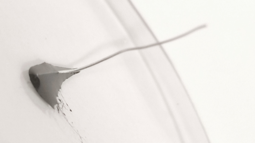
An enzymatic biosensor on a platinum wire.
Biosensors are a new type of sensor that detects specific substances, enabling highly sensitive and selective detection of analytes such as glucose, lactate, and cholesterol. As a promising healthcare technology, they offer real-time, non-invasive monitoring (of blood glucose, for instance) and the resulting wearable data helps health and sports professionals build accurate treatment plans and preventive prescriptions. This tutorial walks through building a proof-of-concept enzymatic lactate biosensor, a promising way to monitor muscle fatigue in athletes.
Context
Biosensors are analytical devices that use biological recognition elements to detect and quantify target molecules. The first one was developed in the 1960s by Clark and Lyons, who measured blood glucose for the first time[2]. Since then, biosensors have advanced considerably and spread across healthcare, food safety, environmental monitoring, and drug discovery[3].
After glucose, lactate is the next biomarker likely to be monitored effectively in the coming years. In vivo human studies show clear links between muscle lactate and muscle fatigue[4][5], and muscle lactate is a well-known marker of exercise-induced fatigue[6][7]. Developing non- or minimally-invasive biosensors for continuous lactate monitoring has recently drawn strong interest in wellness and sports[8], since lactate tracking helps athletes tailor their training to their performance[9][10].
Skills & Opportunities
Biosensing research is multidisciplinary, spanning chemistry, biology, and engineering. This tutorial teaches two main skills.
The first is electrochemistry, the study of the relationship between electricity and chemical reactions. The goal here is to measure an electrical signal that mirrors a chemical reaction, while picking up general lab techniques along the way.
The second is electronic programming. The tutorial uses a programmable analog front-end (AFE) board, the LMP91000, which provides a complete signal path between a sensor and a microcontroller and outputs a voltage proportional to the cell current — a first hands-on approach to an engineering development board.
Requirements
This project requires the following:
- Basic chemical knowledge
- Notions in C++
- An LMP91000 AFE from Texas Instruments
- Lactate Oxidase (LOx)
- PolyPyrrol (PPy)
- Sodium Dodecyl Sulfate (SDS), optional
- A coil of pure platinum wire
- Lactic acid solution
- Phosphate Buffer Saline (PBS)
- Soda
- A magnetic stirrer
- A 1 ml electronic micropipette
- A wash bottle
- A 2 ml Eppendorf tube
- One 500 ml beaker
- Crocodile clips
- Iron wires for electronics
Biosensor Principles
Immobilization Matrix
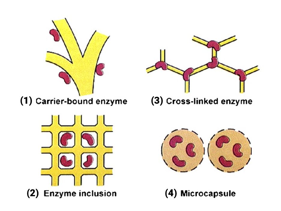
Functionalizing a biosensor means immobilizing the enzyme on a transducer surface[11]. The four main methods are (1) non-covalent adsorption and deposition, (2) physical entrapment, (3) covalent attachment, and (4) bio-conjugation. This tutorial uses physical entrapment, including the enzyme within a polymer network[12].
Enzyme
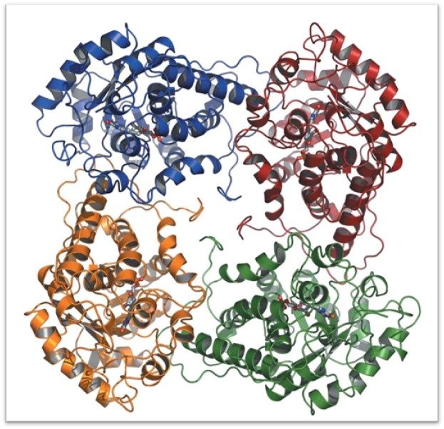
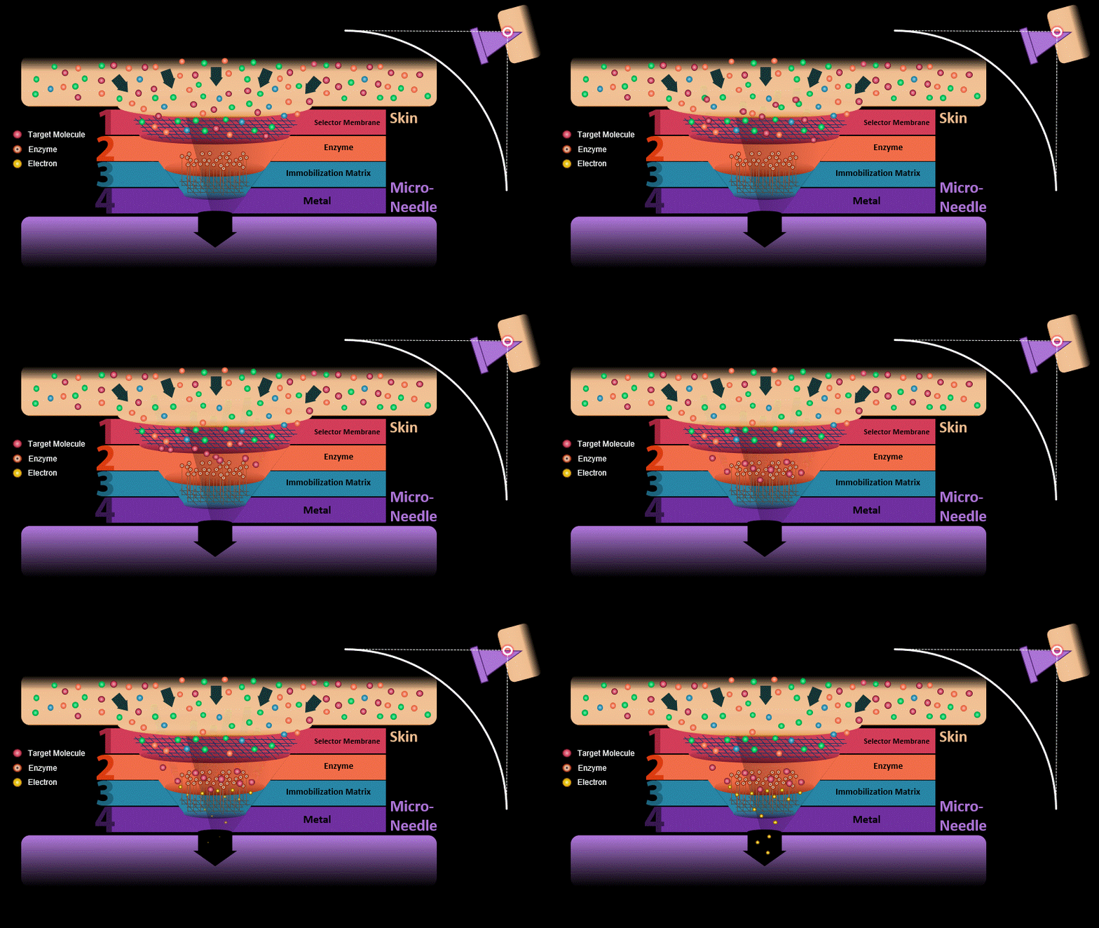
The most common recognition elements for L-lactate biosensors are lactate dehydrogenase (LDH) and lactate oxidase (LOx)[13]. The enzyme catalyzes the oxidation of lactate into pyruvate in dissolved oxygen, producing hydrogen peroxide. Being electrochemically active, the hydrogen peroxide can be reduced or oxidized to yield a current proportional to the lactate concentration[14]. Both enzymes involve simple reactions and allow for a fairly simple sensor design[15]; this tutorial uses LOx for its lower cost.
Membrane
The outer selector membrane has two roles:
- filtering out interferents, biomolecules that could interact with the enzyme and distort the signal;
- regulating the concentration of the target molecule reaching the enzyme, so the biosensor does not run short of oxygen and saturate[16].
This tutorial skips membrane design: its two functions are not required for in vitro testing, and the membrane also tends to reduce the biosensor's sensitivity.
Tutorial
1. Biosensor Functionalization
1.1. Prepare the platinum wire. Cut the platinum wire to a 5-centimeter length.
1.2. Prepare the PBS solution. Dilute PBS powder in 1 liter of distilled water and fill a wash bottle with it.
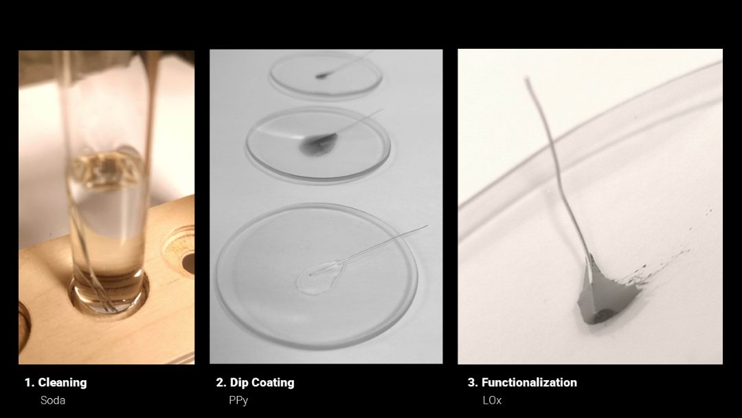
1.3. Clean the platinum wire. Dissolve 0.8 g of solid soda in a 100 ml beaker of distilled water with a stirrer. Leave the wires in the soda solution for 10 minutes, remove them, clean thoroughly with ethanol, and dry at room temperature.
1.4. Prepare the polypyrrole solution. Weigh 0.4 g of PPy powder into a beaker with 20 ml of acetone. Stir for 30 minutes at room temperature until fully dissolved, then add 80 ml of distilled water and stir for 30 more minutes to obtain a homogeneous PPy solution.
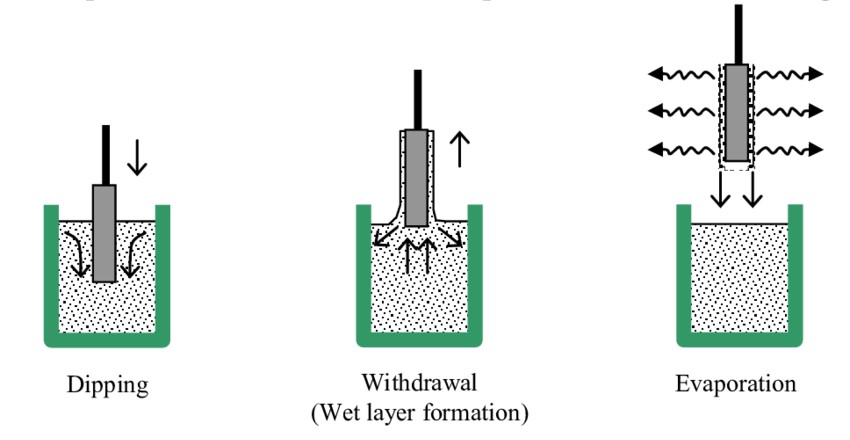
1.5. Dip-coat the platinum wire with PPy. Dip the cleaned wire into the PPy solution and withdraw it slowly at about 2 cm/min for 2 minutes (use a stopwatch and ruler if you have no dip coater, or leave it immersed motionless). Dry the wire at 60 °C in an oven for 30 minutes.
1.6. Prepare the functionalization solution. Pour 2 µl of LOx into a 2 ml Eppendorf tube with 2 ml of PBS using a micropipette. Close and shake vigorously to homogenize.
1.7. Immobilize lactate oxidase by entrapment. Pour 0.2 ml of the functionalization solution onto the last 2 cm of the PPy-coated wire to immerse it. Leave for 6–12 hours at room temperature; the enzyme becomes entrapped within the PPy layer.
1.8. Wash the immobilized wire. Remove the wire from the LOx solution and rinse it with PBS.
2. LMP91000 Programming
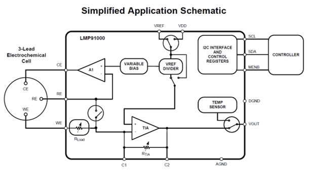
Electrochemical biosensor tests require a potentiostat — an electronic circuit that applies a potential to a working electrode (WE)[23]. By applying the molecule's oxidation potential (+650 mV for lactate), the enzyme catalyzes its oxidation, producing one or more electrons. The resulting current flows through the WE to the circuitry and a counter electrode (CE), where op-amps amplify it to the microampere range. Chronoamperometry (CA) applies a fixed potential at the WE and measures current over time[24], usually needing about an hour of calibration until the WE–CE potential stabilizes. The current is proportional to the lactate concentration at the WE surface. The LMP91000 is a popular potentiostat board for micro-power electrochemical sensing[25].
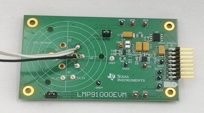
2.1. Prepare the electrodes. Solder three wires to the CE, WE, and RE.
2.2. Wiring. Connect 3V3, GND, SDA, and CLK between the LMP and the WeMos. Using an alligator clip, connect the LMP's Vout pin to the WeMos's A0 pin.
2.3. Download the code. Get the code from the BioWatch GitHub, run the chronoamperometry code, and check with a potentiometer that the board works.
| # | Top Pins | # | Bottom Pins |
|---|---|---|---|
| 15 | 16 | SCS2 | |
| 13 | GPSI 3.3V | 14 | GPSI 5V |
| 11 | SDA | 12 | SCL |
| 9 | CLK | 10 | GND |
| 7 | MOSI | 8 | MEMB INT |
| 5 | MISO | 6 | DEV INT |
| 3 | SCK | 4 | GND |
| 1 | SCS1 | 2 | GND |
LMP91000 pinout.
3. Experimentation
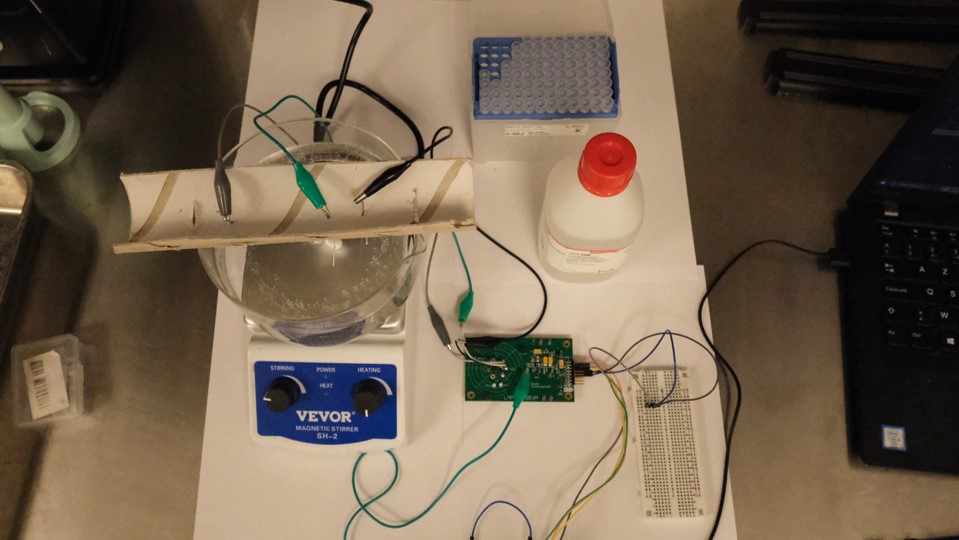
3.1. Connect the electrodes. Connect the functionalized platinum wire to the WE of the LMP91000 with an alligator clip. Cut and connect two more platinum wires to the CE and RE the same way, then check the connections with a voltmeter.
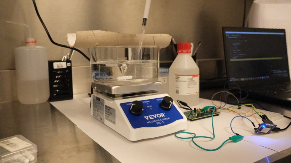
3.2. Set up the electrochemical cell. Fill a clean beaker with 400 ml of PBS and stir it constantly at medium speed with a magnetic stirrer.
3.3. Place the electrodes. Insert the platinum wires into the cell and start the chronoamperometry code. Check that the values are consistent, then wait 3600 seconds.
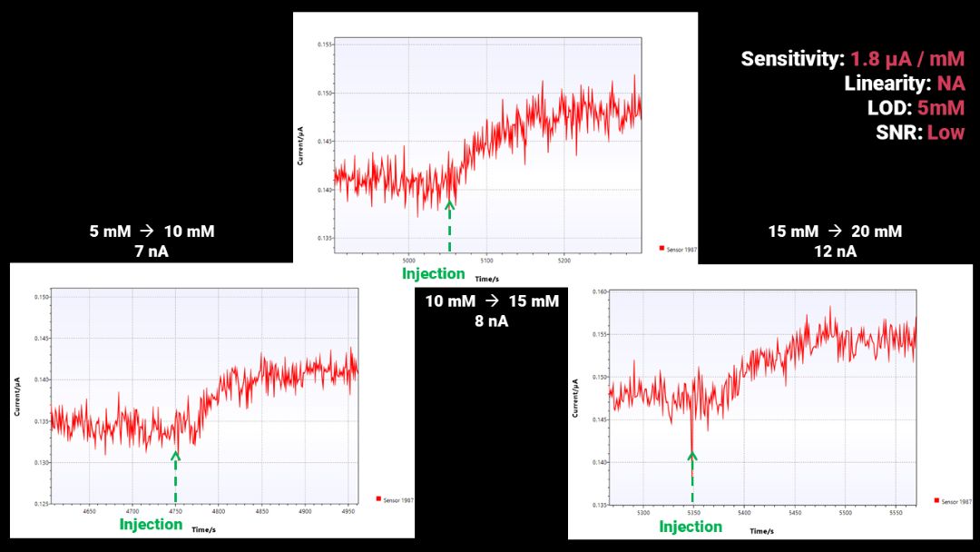
3.4. Inject lactate. Add 160 µl of lactic acid with a propette to reach a 4 mM solution. Repeat 4 times every 5 minutes, up to 20 mM.
3.5. Exploit the results. Copy the data from the console and paste it into a spreadsheet. Split time and current into two columns using the "," separator, and convert full stops into commas.
3.6. Characterize the sensor. Plot the inputs against time, identify the injections, and compute the sensor's sensitivity and linearity.
Conclusion
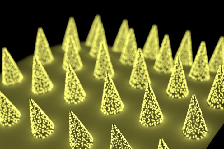
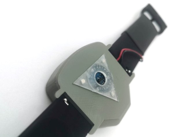
This tutorial designs a simple lactate biosensor from scratch using the most accessible equipment possible. The sensors built and tested perform poorly, likely because the non-optimized wiring lets in strong signal perturbations. Future work will test them with a commercial potentiostat. The platinum wire could also be replaced by micro-needles coated with platinum paste, which could then be integrated into the BioWatch.
References
- Sterling J. Novel Microneedle Patch on the Skin Can Test for Biomarkers. GEN — Genetic Engineering and Biotechnology News. 2021. Link
- Yoo EH, Lee SY. Glucose biosensors: an overview of use in clinical practice. Sensors (Basel). 2010. doi:10.3390/s100504558
- Tetyana P, et al. Biosensors: Design, Development and Applications. Nanopores, IntechOpen. 2021. doi:10.5772/intechopen.97576
- Cairns SP. Lactic Acid and Exercise Performance. Sports Med. 2006. doi:10.2165/00007256-200636040-00001
- Messonnier L, Dubouchaud H. Le lactate : sa cinétique, son métabolisme… Movement & Sport Sciences. 2010.
- Finsterer J. Biomarkers of peripheral muscle fatigue during exercise. BMC Musculoskeletal Disorders. 2012. doi:10.1186/1471-2474-13-218
- Wan JJ, et al. Muscle fatigue: general understanding and treatment. Exp Mol Med. 2017. doi:10.1038/emm.2017.194
- Chien MN, et al. Continuous Lactate Monitoring System Based on Percutaneous Microneedle Array. Sensors. 2022. doi:10.3390/s22041468
- Billat LV. Use of blood lactate measurements for prediction of exercise performance. Sports Med. 1996. doi:10.2165/00007256-199622030-00003
- Goodwin ML, et al. Blood lactate measurements and analysis during exercise. J Diabetes Sci Technol. 2007. doi:10.1177/193229680700100414
- Nguyen HH, et al. Immobilized Enzymes in Biosensor Applications. Materials (Basel). 2019. doi:10.3390/ma12010121
- Homaei AA, et al. Enzyme immobilization: an update. J Chem Biol. 2013. doi:10.1007/s12154-013-0102-9
- Rathee K, et al. Biosensors based on electrochemical lactate detection: A comprehensive review. Biochem Biophys Rep. 2016. doi:10.1016/j.bbrep.2015.11.010
- Meyerhoff C, et al. On-line continuous monitoring of blood lactate by a wearable enzymatic amperometric sensor. Biosens Bioelectron. 1993. doi:10.1016/0956-5663(93)80025-k
- Rathee K, et al. Biosensors based on electrochemical lactate detection: A comprehensive review. Biochem Biophys Rep. 2015. doi:10.1016/j.bbrep.2015.11.010
- Davies ML, et al. Polymer membranes in clinical sensor applications. I. Biomaterials. 1992. doi:10.1016/0142-9612(92)90147-g
- Yu H, et al. Recent Progress of Platinum Nanoparticle-Based Electrochemistry Biosensors. Front Chem. 2021. doi:10.3389/fchem.2021.677876
- Chu X, et al. Amperometric glucose biosensor based on platinum nanoparticles and carbon nanotube electrode. Talanta. 2007. doi:10.1016/j.talanta.2006.09.013
- Jain R, Jadon N, Pawaiya A. Polypyrrole based next generation electrochemical sensors and biosensors: A review. TrAC Trends in Analytical Chemistry. 2017. doi:10.1016/j.trac.2017.10.009
- Gabriela P, Bizerea O, Vlad-Oros B. Sol-gel technology in enzymatic electrochemical biosensors for clinical analysis. 2011. doi:10.5772/19622
- Dip Coating: Practical Guide to Theory and Troubleshooting. Ossila. 2023. Link
- Sanchez-Herencia AJ. Water Based Colloidal Processing of Ceramic Laminates. Key Engineering Materials. 2007. doi:10.4028/www.scientific.net/kem.333.39
- Potentiostat: a simple and short explanation. PalmSens. 2023. Link
- Electro-analytical techniques. Zimmer and Peacock. 2023. Link
- Mouser Electronics. Potentiostat AFE configurable LMP91000 TI. 2022. Link
- LMP91000. Zimmer and Peacock. 2023. Link